
(HealthDay)—Joint recesses with bone erosion are more likely to exhibit greater severity of joint inflammation on ultrasound (US) examination, according to a study published online Oct. 25 in the Journal of Clinical Ultrasound.
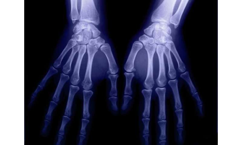
York Kiat Tan, from Singapore General Hospital, and colleagues used an extended 36-joint sonographic examination in 30 patients with rheumatoid arthritis to identify joints commonly exhibiting bone erosion. A combined US score (CUS) was calculated by summing power Doppler (PD) and gray-scale (GS) joint inflammation scores (semi-quantitative [0 to 3] grading) at each joint recess.
The researchers observed bone erosion in 144 of 1,080 joints (13.3 percent) and 189 of 1,800 joint recesses (10.5 percent). The wrist (34 percent of 144), first metatarsophalangeal joint (13.2 percent), thumb interphalangeal joint (9 percent), second metacarpophalangeal joint (MCPJ; 7.6 percent), and third MCPJ (7.6 percent) were the joints most frequently associated with bone erosion. With and without bone erosion, mean US scores for joint recesses were a PD of 0.36 versus 0.01; GS of 1.77 versus 0.47; and CUS of 2.13 versus 0.49.
Source: Read Full Article