Exercising regularly is one of the best defenses against metabolic diseases, such as obesity and diabetes—but why? It’s a question that scientists are still struggling to answer. While exercising changes the molecular behavior of muscles, it’s not well understood how these molecular changes improve metabolic health.
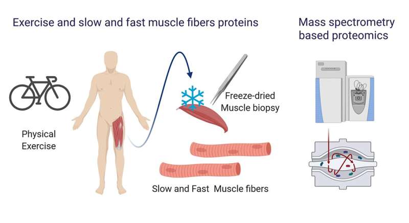
Scientists at the University of Copenhagen have now developed a new technology that allows researchers to study muscle biology on a more detailed level—and hopefully find some new answers. They extracted ‘fast’ and ‘slow’ twitch muscle fibers from freeze-dried muscle samples that were taken before and after 12 weeks of cycling exercise training. Their comprehensive analysis of the protein expression of the fibers provides new evidence that the fiber types respond differently to exercise training.
The research, which was published in Nature Communications, also demonstrates the untapped potential of freeze-dried samples that are located in freezers around the world.
“Metabolic disorders and several muscle diseases are known to affect or spare specific fiber types, therefore detailed investigation of specific fiber types is crucial. Previous studies involving large-scale protein analysis of muscle fibers required isolation of single muscle fibers from freshly obtained muscle biopsies. Since isolation of muscle fiber is time consuming, this approach has its inherent limitations. Our method enables muscle fiber analysis of already collected muscle biopsies as well as paves the new way for future studies,” says Associate Professor Atul Deshmukh from the Novo Nordisk Foundation Center for Basic Metabolic Research (CBMR) at the University of Copenhagen.
Messy and confusing muscle samples
Skeletal muscle contains tiny fibers that can be categorized as either fast or slow twitch. Simply put, fast twitch fibers create explosive energy but get tired quickly, while slow twitch fibers are less energetic but have more endurance. Most people have an even number of both types in their muscles, but the ratio can vary widely between people. This means that exercise may benefit people differently, depending on this ratio.
In a muscle, thousands of fibers are bundled together by connective tissue and interspersed with a range of cell types with a supporting function. Because of all these different cell types, scientists can struggle to interpret the results of a whole muscle sample, and connect observed changes to specific cell types.
Understanding the potential of studying individual fibers, Atul Deshmukh teamed up with Professor Matthias Mann from the Novo Nordisk Foundation Center for Protein Research, and the Wojtaszewski Group from the Department of Nutrition, Exercise and Sports, both at the University of Copenhagen.
Honing in on muscle fibers
They recruited healthy individuals for 12 weeks of endurance exercise training and collected muscle samples before and after exercise, which were then freeze-dried. They then extracted fast and slow twitch muscle fibers from the samples and carried out high-resolution mass spectometry-based proteomics—a tool that allows scientists to measure thousands of proteins simultaneously in the different samples.
They identified more than 4,000 different proteins in the samples, and discovered that exercise training changed the expression of hundreds of different proteins in both fast and slow twitch fiber-types. Importantly, they discovered differences to the protein expression of the two fiber-types after exercise, which demonstrates that fast and slow twitch muscle respond differently.
“Our method can be scaled for high-throughput analysis of hundreds of individual muscle fibers from a single biopsy. Coupling this approach with the modern high sensitivity mass spectrometer may help to understand the fiber type heterogeneity in healthy and diseased skeletal muscle,” says Associate Professor Atul Deshmukh from the Novo Nordisk Foundation Center for Basic Metabolic Research (CBMR) at the University of Copenhagen.
Drugs that don’t accidentally target the heart
By mass, skeletal muscle is the largest organ in the body and even small changes can have tremendous impact on whole body metabolism. Skeletal muscle is therefore an interesting pharmacological target tissue with great potential in treatment of metabolic diseases. One challenge is however to avoid side effects in heart muscle, for example, which consists of specialized fibers with some similarities to slow twitch skeletal muscle fibers.
Source: Read Full Article
