The study covered in this summary was published on researchsquare.com as a preprint and has not yet been peer reviewed.
Key Takeaway
-
Adding contrast imaging to standard 2D/3D digital mammography improves detection of recurrent or new malignancies in patients with a history of breast cancer.
Why This Matters
-
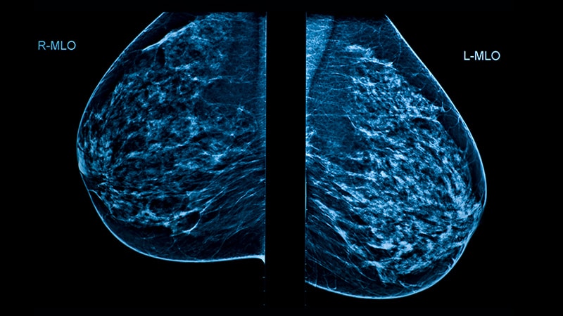
Women with a history of breast cancer or ductal carcinoma in situ (DCIS) undergo routine surveillance for new or recurrent disease.
-
Ultrasound or MRI are often used to supplement digital mammography.
-
Contrast-enhanced mammography has higher sensitivity than mammography and ultrasound as well as better accessibility than MRI.
-
The findings support use of contrast-enhanced mammography for routine surveillance in breast cancer survivors.
Study Design
-
In 2018, the Royal Melbourne Hospital in Australia switched to contrast enhanced mammography, in combination with 2D/3D mammography, for routine surveillance of patients with a history of breast cancer or DCIS, replacing the previous standard of 2D/3D mammography with selective ultrasound.
-
Investigators reported the outcomes, comparing contrast enhanced mammography with digital mammography.
Key results
-
Overall, 73 of 1191 patients (6.1%) were recalled for further assessment.
-
Almost half of recalls (35 patients) were true positives — 26 invasive cancers and 9 cases of DCIS; the other half (38 patients) were false positives resulting in a positive predictive value 47.9%
-
Of the 73 recalled patients, 9 lesions were seen on 2D/3D mammography alone; 23 lesions were identified using both 2D/3D mammography and contrast— among these, 83% were true positives.
-
According to the researchers, 41 of 66 biopsied recalls “were unlikely to have been identified had contrast not been used.”
-
Contrast-only true positives were found in patients with both low and high breast density.
-
Introduction of contrast-enhanced mammography led to a steady decrease in the use of whole breast ultrasound for surveillance.
Limitations
-
False negative rates couldn’t be determined.
-
Patients were offered surveillance contrast-enhanced mammography at various times after their initial cancer treatment.
-
It is possible that some lesions found on surveillance contrast- enhanced mammography were present at the time of the initial cancer diagnosis before the widespread use of contrast.
Disclosures
-
No funding source was reported.
-
One author served once on an advisory board for Hologic, and another reported research funding from the company.
This is a summary of a preprint research study, “Contrast Enhanced Mammography in Breast Cancer Surveillance,” led by Kenneth Elder, MSc, MPhil, BMBS, of the Royal Melbourne Hospital, Melbourne, Australia, provided to you by Medscape. The study has not been peer reviewed. The full text can be found at researchsquare.com.
M. Alexander Otto is a physician assistant with a master’s degree in medical science and a journalism degree from Newhouse. He is an award-winning medical journalist who has worked for several major news outlets before joining Medscape and also an MIT Knight Science Journalism fellow. Email: [email protected].
For more from Medscape Oncology, join us on Twitter and Facebook
Source: Read Full Article