Self-reported maternal psychological distress during the COVID-19 pandemic may be associated with changes in the brain of developing fetuses, according to a study published in Communications Medicine. The study involved 65 women who were pregnant during the pandemic (June 2020 to April 2021) and 137 who were pregnant prior to the pandemic (March 2014 to February 2020). None of the participants assessed during the course of the pandemic were known to have been infected with SARS-CoV-2, the virus that causes COVID-19. The study assessed the potential impact of the pandemic itself on pregnant women and their developing fetuses before birth, rather than the impact of COVID-19 infections.
Catherine Limperopoulos and colleagues imaged the brains of fetuses in the wombs of mothers who were pregnant before and during the COVID-19 pandemic using Magnetic Resonance Imaging (MRI). The imaging assessed the surface structure of the brain, including cortical folding and the wrinkled shape (gyrification) and depth of the wrinkles (sulcal depth) of the brain surface.
From the 202 participants, authors asked 173 mothers questions to investigate any distress experienced during pregnancy including anxiety, stress and depression. The authors found stress and depression were reported proportionally more in the mothers who were pregnant during the pandemic. Overall, 34 (27.6%) women in the pre-pandemic cohort and 26 (52.0%) women pregnant during the pandemic were considered to have high psychological distress. Anxiety levels stayed consistent across groups.
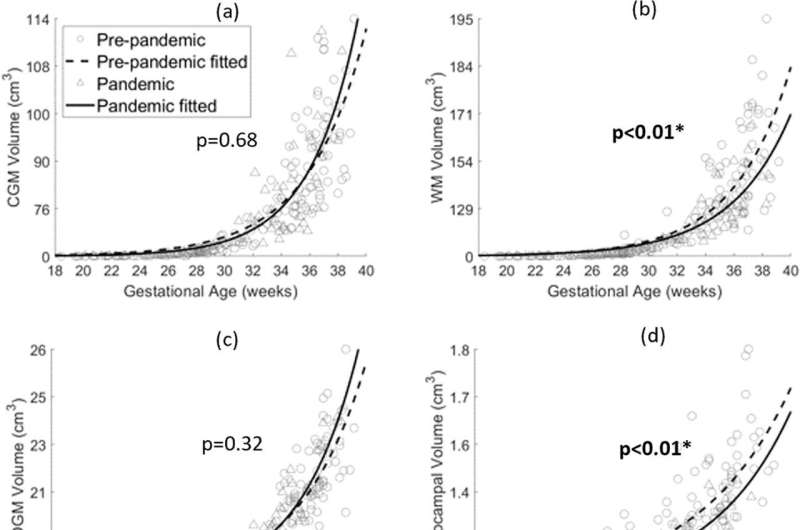
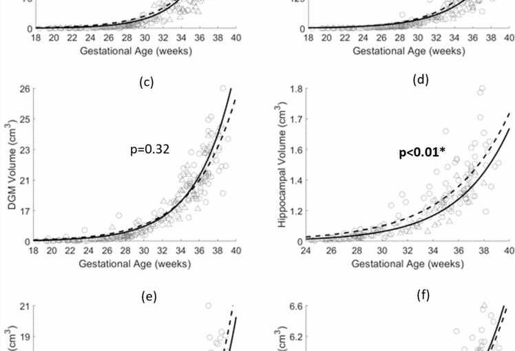
The authors observed that three brain structure and volumetric measures (fetal white matter of the cerebrum, hippocampal, and cerebellar volumes) were decreased in the fetuses from the pandemic cohort compared to the pre-pandemic cohort. Development in these brain structures was negatively associated with anxiety, stress and depression scores. When also including mothers reporting low stress in the analysis, the authors observed fetuses of pregnant women in the low stress group had lower volumes across the three brain measures in the pandemic cohort compared to the pre-pandemic cohort. The authors suggest this variability and inconsistency indicates multiple factors are involved in fetal brain development.
When observing brain structure, the authors found cortical surface area and local gyrification index were decreased in all four lobes, while sulcal depth was lower in the left frontal, parietal, and occipital lobes in the pandemic cohort. The authors suggest these metrics may indicate delayed brain gyrification.
Source: Read Full Article
