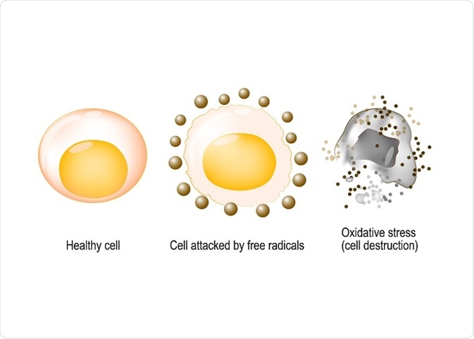
Oxidants, such as reactive oxygen species (ROS), are known to induce damage to therapeutic proteins. Although cells and tissues usually have antioxidant systems to combat ROS and maintain the balance between formation and neutralization of ROS, in some cases this delicate balance may be perturbed, leading to physiological and molecular damage to proteins. In these cases, methods need to employed to reduce the likelihood of damage.
 Designua | Shutterstock
Types of oxidative damage
Oxidation may be induced by the modification of a protein directly using ROS, or during a reaction with the by-products of oxidative stress. Oxidative agents that can induce oxidative damage include H2O2, HOCl or xenobiotics, such as paraquat, acetaminophen, cigarette smoke, metals like Fe2+, Cu+, gamma radiations, ultraviolet light, ozone, etc.
Two amino acids that are highly prone to oxidation are cysteine and methionine. Oxidation in cysteine forms disulfide bonds and thiyl radicals, while oxidation in methionine forms methionine sulfoxide. Protein-bound metal ions are also potent oxidants that are able to reduce hydrogen peroxide to form a reactive intermediate near amino acid side chains. Indirect oxidation of proteins may also occur by addition of oxidatively modified lipids, amino acids and sugars.
What are the consequences of oxidation of therapeutic proteins?
Oxidation can lead to inhibition of enzyme activities of the proteins; furthermore, it can also exert effects on a cell or systemic metabolism based on the degree of modification. Studies have also shown that several proteins are oxidized during the process of aging.
In many cases, oxidation inactivates a protein. For example, oxidation of the plasma protein, fibrinogen, can result in the loss of its clot-forming ability. This can have serious health implications, as one of the causes of rheumatoid arthritis is oxidation of synovial fluid immunoglobulins.
Minimizing oxidation and oxidative damage to proteins
Oxidative changes in proteins can have large repercussions where it can affect the bioactivity and pharmacokinetic parameters of the therapeutic proteins. Thus, methods to reduce the oxidation are often introduced.
Lowering oxygen tension is one way how to achieve that goal. Studies show that oxidation often occurs in the periplasmic space of E.coli which leads to oxidation of proteins, such as methionine. However, when the cells were grown in anaerobic conditions, oxidation of proteins was not observed.
Using pH that minimizes oxidations is another way. In some cases, the preparation of therapeutic proteins was carried out under inert gases, or certain pH was used that minimises oxidation and reduction reactions. For example, conducting an experiment at acidic pH can minimise the reactivity of sulfhydryl.
Studies have also tried to remove the variants of a protein that are oxidized during a reaction by employing downstream purification methods. For example, during the isolation of human insulin-like growth factor, several variants are present that consist of the methionine-sulfoxide variant. This version of protein folds improperly and is ineffective. However, this variant can be removed using reverse-phase high-performance liquid chromatography, and optimizing pH, ionic strength, and temperature as a downstream process.
Can antioxidants be used to minimize oxidative damage?
The recombinant monoclonal antibodies (such as HER2) undergo oxidation when exposed to light or increased temperatures. The primary site of oxidation is methionine in the Fc region of the antibody; moreover, the levels of oxidation were increased by the presence of NaCl.
Addition of antioxidants, such as methionine, sodium thiosulfate, catalase or platinum, could act as an oxygen scavenger or free radical and, in turn, prevent the oxidation of methionine. Thus, the temperature-induced oxidation of protein could be abrogated by specific amounts of methionine and thiosulfate, as evidenced by recent studies.
Sources
- pdfs.semanticscholar.org/6b37/1c4976afce27d59cd76bbf487a69286623e6.pdf
- https://www.ncbi.nlm.nih.gov/pubmed/9383735
- openaccess.leidenuniv.nl/…/02.pdf?sequence=13
Last Updated: Mar 27, 2019

Written by
Dr. Surat P
Dr. Surat graduated with a Ph.D. in Cell Biology and Mechanobiology from the Tata Institute of Fundamental Research (Mumbai, India) in 2016. Prior to her Ph.D., Surat studied for a Bachelor of Science (B.Sc.) degree in Zoology, during which she was the recipient of anIndian Academy of SciencesSummer Fellowship to study the proteins involved in AIDs. She produces feature articles on a wide range of topics, such as medical ethics, data manipulation, pseudoscience and superstition, education, and human evolution. She is passionate about science communication and writes articles covering all areas of the life sciences.
Source: Read Full Article
