About 600 corneal transplantations are performed in Denmark every year. Most of them are performed as treatment of the eye disease “Fuchs endothelial dystrophy,” which causes cellular layers on the back of the cornea to become unclear, resulting in severely impaired vision. In Denmark, the disorder is treated surgically with a corneal transplantation in which the diseased cellular layer on the back of the cornea is removed and replaced by donor tissue. The current method for securing the attachment of donor tissue on the back of the cornea is facilitated by injecting a small air bubble that pushes the donor tissue towards the patient’s cornea. However, a completely different solution may be better.
The eye specialists at the Department of Ophthalmology at Rigshospitalet—who are conducting research into new techniques for corneal transplantation—would like to have greater knowledge about precisely this question. However, they experience that there is no usable solution available when new techniques for surgeries on humans are to be developed.
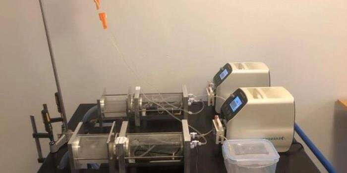
“We approached DTU Mechanical Engineering’s product development department to inquire whether we could jointly develop a model of the anterior chamber of the eye that made it possible to conduct research into how a human cornea thrives best after a transplantation,” says Javier Cabrerizo, Eye Surgeon and Clinical Associate Professor at the department.
Solution to several challenges
It was not a simple assignment, but an obvious challenge for the researchers at DTU, who work precisely with designing things with multiple functions.
“The model should first and foremost mimic the pressure and temperature found in the anterior chamber of the eye. In addition, the flow of fluid to the back of the cornea and tear film located on the front of the cornea had to be mimicked. Finally, the model was to support the post-operative phase, where the patient first lies down and later sits and stands up,” explains Nicklas Werge Svendsen, DTU Mechanical Engineering, who has been responsible for developing the model.
The goal was to develop a model that could keep a cornea alive for six days.
“We succeeded in this, which provides us with sufficient time for observing how the cells on the posterior layer of the cornea react after a transplantation,” says Javier Cabrerizo.
New theoretical basis
As a derived effect of the work to construct the eye model, Nicklas Werge Svendsen has expanded the theoretical basis for handling design problems where the solution principles of multiple functions influence each other. This is of particular interest to that part of the design research that is inspired by solutions in nature’s plants, animals or fungi —also called biologically inspired design.
“Based on my work with developing the model of the anterior chamber of the eye, I’ve created an approach to the theoretical design work that—using modeling of functions and means—makes it possible to see new and different relationships than before,” says Nicklas Werge Svendsen.
The extension of the theoretical basis for modeling of functions and means will be used in design work in the future. However, Nicklas Werge Svendsen is particularly proud of having produced a practical model that can be used in research into corneal disorders. That joy is shared by Rigshospitalet’s Department of Ophthalmology.
Source: Read Full Article
