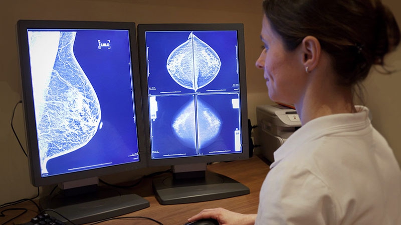
The study covered in this summary was published on ssrn.com as a preprint and has not yet been peer reviewed.
Key Takeaway
-
Artificial intelligence (AI)–assisted mammography substantially reduces the workload of standard double reading by two radiologists and improves cancer detection without increasing false positives findings, according to a randomized trial that included more than 80,000 women.
Why This Matters
-
Retrospective studies have shown that AI improves mammography screening accuracy while reducing screen-reading workload.
-
The new study is the first prospective, randomized trial to investigate the use of AI for mammography screening.
-
The results confirm the benefits of AI in mammography and strongly support implementing it into screening programs.
Study Design
-
The Mammography Screening With Artificial Intelligence (MASAI) trial randomly assigned 40,003 women to AI-supported mammography and 40,030 to standard, double-reading mammography without AI.
-
Women were aged 40 to 74 years and were routinely undergoing screening every 1 to 2 years on the basis of risk as part of Sweden’s population-based mammography program. There were no exclusion criteria.
-
In the experimental arm, the AI system (Transpara version 1.7.0, Screenpoint Medical) generated a malignancy risk score that was based on its assessment of screening images.
-
Women with lower scores were triaged to review by a single radiologist. Women with higher scores were triaged to double reading; radiologists made the decision as to whether to recall women for further investigation.
-
Radiologists in the AI arm had access to the system’s risk scores and computer-aided detection marks.
-
Overall, 15 of 16 breast radiologists in the study had more than 2 years of experience, and 14 had more than 5 years. For 12 radiologists, the yearly reading volume was at least 5000 cases.
-
The trial used the Senographe Pristina 3D mammography system from GE Healthcare.
Key Results
-
AI-supported screening detected 41 more cancers, a 20% increase over standard double reading.
-
Overall, 19 of the 41 cancers were invasive, and 22 were in situ.
-
The cancer detection rate was 6.1/1000 women in the AI group, vs 5.1/1000 with standard care.
-
The recall rate was 2.2% with AI and 2% without; the rate of false positive findings was 1.5% in both arms.
-
Radiologists conducted 36,886 fewer readings in the AI arm, a 44% drop in screen reading workload.
Limitations
-
There is a question of generalizability in practices that use different AI and mammography systems and that have less experienced radiologists.
-
The interval-cancer rate, which could affect false positive results, will not be known until the full study population of 100,000 women have had at least 2 years of follow-up (by December 2024).
-
The relatively higher detection of in situ cancers with AI raises the possibility of overdiagnosis.
Disclosures
-
The work was funded by the Swedish Cancer Society and others.
-
The study’s lead is an advisor for Siemens Healthineers and has received lecture honorarium from AstraZeneca. Another investigator is a researcher for Screenpoint Medical.
This is a summary of a preprint research study, “The Safety of an Artificial Intelligence Supported Screen-Reading Procedure Versus Standard Double Reading in the Mammography Screening with Artificial Intelligence (MASAI) Trial: A Randomised, Controlled, Screening Accuracy Study,” led by Kristina Lang of Lund University, Sweden, provided to you by Medscape. The study has not been peer reviewed. The full text can be found at ssrn.com.
M. Alexander Otto is a physician assistant with a master’s degree in medical science and a journalism degree from Newhouse. He is an award-winning medical journalist who has worked for several major news outlets before joining Medscape and also an MIT Knight Science Journalism fellow. Email: [email protected].
For more from Medscape Oncology, join us on Twitter and Facebook.
Source: Read Full Article
